The viability of a cell or population of cells is an overall measure of the health of the cells. Viability assays are designed to assess the physical and metabolic state of the cell in order to determine the impact of treatment or culture conditions on cellular homeostasis.
The critical link between cellular health and disease underscores the necessity for assays that can measure cell viability in different experimental contexts and model systems.
Cell viability is measured using a variety of techniques and experimental platforms. Some techniques, such as enzyme-linked immunosorbent assay (ELISA) based viability assays, utilize spectral-based microplate readers for analysis. Other commonly used assays employ immunofluorescence, flow cytometry, or western blotting as a readout.
Cell viability vs cell proliferation assays:
Multiple techniques are used for measuring cell viability. To confirm experimental outcomes, multiple independent assays should be performed. Techniques for analyzing cell viability include:
| Assay | What is Measured | |
|---|---|---|
| Detect metabolic activity from viable cells with active mitochondria | ||

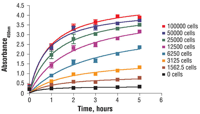
XTT Cell Viability Kit #9095: C2C12 cells were seeded at varying density in a 96-well plate and incubated overnight. The XTT assay solution was added to the plate and cells were incubated. The absorbance at 450 nm was measured at 1.0, 2.0, 3.0, 4.0, and 5.0 hours. |
||
|
7-AAD/CFSE Cell-Mediated Cytotoxicity Assay Kit Trypan blue |
Cell viability dyes are used to monitor dye uptake into nonviable cells; the dyes are excluded from entering viable cells | |

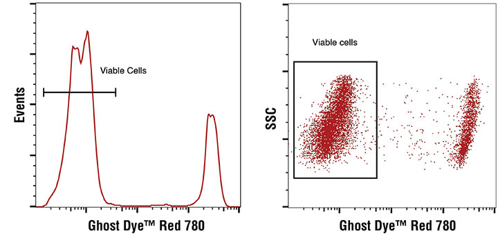
Ghost Dye™ Red 780 Viability Dye #18452: Flow cytometric analysis of live and fixed/permeabilized human peripheral blood mononuclear cells, combined and stained with Ghost Dye™ Red 780 Viability Dye. Viable gate is indicated. |
||
|
Phospho-histone H3 (western blot) Proliferating cell nuclear antigen (PCNA) Ki-67 (IHC) |
Measure expression levels of proteins required for (or involved in) cell division. | |

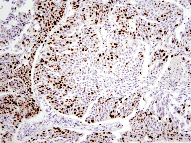
Ki-67 (D2H10) Rabbit mAb (IHC Specific) #9027: Immunohistochemical analysis of paraffin-embedded human colon carcinoma using Ki-67 (D2H10) Rabbit mAb (IHC Specific). |
||